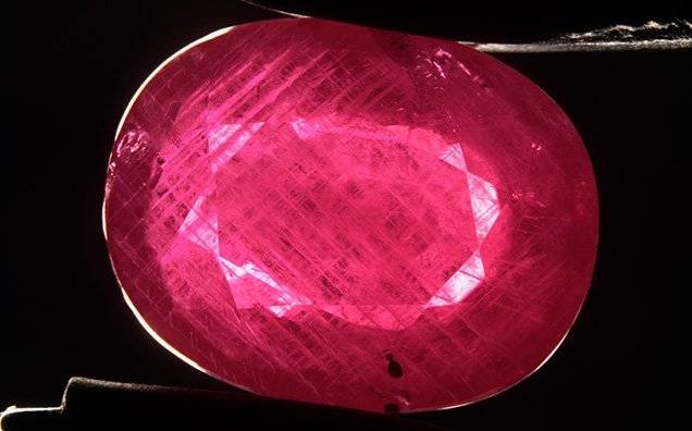
Rubies and pink sapphire from Greenland are a new addition to the trade. The deposits have been known for decades, but only in the last three years have the finished gems become available in the market. The vast majority of the material is mined by the company Greenland Ruby and disclosed as treated. The habits of the rough corundum crystals are tabular, barrel, or rounded in shape. Their color ranges from dark red to very light pink, with a varying purplish or grayish cast.
Since the stones are mined from a primary deposit, they are often attached to other minerals. Most are mechanically removed at the mine site during the highly automated sorting process. At Greenland Ruby’s treatment facility, some stones might be further manually trimmed or clipped to remove more of the matrix. Next, the rubies and pink sapphires are intensely cleaned with strong acids. These dissolve most minerals but do not affect the corundum. Lime is used to neutralize the acid, sometimes leaving a thin white coating on the rough stones.
After cleaning, the stones are heated with flux in crucibles and heated in electric furnaces for multiple hours at high temperatures (>1500°C). This method improves the clarity by healing thin fractures. Excess flux covers the rough stones in a thin glassy layer. After cooling, this product is polished as cabochons or beads. Higher-clarity pieces are faceted. The small amount of corundum produced by artisanal miners is treated in a similar fashion, but their exact procedures are not known.
Inclusion scenes of untreated stones are described in C.P. Smith et al. (“Ruby and pink sapphire from Aappaluttoq, Greenland,” Journal of Gemmology, Vol. 35, No. 4, 2016, pp. 294–306) and K. Thirangoon (“Ruby and pink sapphire from Aappaluttoq, Greenland: Status of ongoing research,” GIA Research News, March 23, 2009). These studies highlight the unique and exotic suite of crystal inclusions, including sapphirine and cordierite. We were able to complement these earlier works by studying some of the high-grade material recently produced by Greenland Ruby that has been treated. This is a very intense treatment procedure that significantly alters the inclusion scenes of Greenland’s rubies and pink sapphires.
The most obvious internal features are linked to the omnipresent twinning in corundum from Greenland (figure 1). The twinning planes themselves are not altered by the treatment. The so-called Rose channels, which are hollow tubes at the intersection of twin planes, do show changes. Small fractures, often healed, develop around the linear features, while the tubes themselves usually take on a more spotted appearance when viewed at higher magnification (figure 2A).
Most crystal inclusions show strong alteration, as expected for high-temperature treatment. This makes the unique inclusion scene of Greenland ruby and sapphire unrecognizable. Many of these inclusions transform into “snowballs” with a white frosty opaque nature, often accompanied by discoid fractures that can show signs of healing (figures 2, B and C). In rare cases, crystal inclusions such as zircon remain unaltered.
Less common in Greenland rubies are particles and platelets. Since they require a correct alignment of the observer, platelet, and light source to be detected, they are often overlooked. The particles are a mix of elongated and irregularly shaped platelets. Some could be described as short, stubby needles resembling arrowheads (figure 3, left). They can also appear in dense clouds of finer particles with a roughly hexagonal outline (figure 3, right). These look very similar to the features observed in East African rubies from Mozambique and Madagascar, where they are very common. These seem to be only subtly affected by the treatment. In rare cases, the platelets take a roughly hexagonal form with concentric brightness zoning, resembling a fried egg “sunny-side up.” These may be reminiscent of inclusions typically associated with Thai rubies, although Thai rubies almost never show the presence of other particles. We suspect that the unique appearance of these platelets is due to the intense heat they have been subjected to.
Apart from the twinning features, the most obvious features in the treated stones are flux-healed fractures. These are caused entirely by the treatment and are diagnostic of flux healing. As such, they are completely unrelated to a specific origin. Remnants of the flux trapped in the healing fracture can appear as small irregular droplets or connecting worm-like tubes (figure 4, A and B). These might bear some resemblance to natural fingerprints, but they usually have a coarser appearance with more translucent whitish patterns. When the healing is incomplete, the flux can be present in the fractures as a glassy substance showing swirling patterns and trapped gas bubbles (figure 4C).
The treatment has no impact on the corundum’s trace element chemistry. The composition of the studied suite is in line with the data reported in Smith et al. (2016). The only values that show some deviation are the titanium concentrations, which are lower in the samples analyzed in this study (12–102 ppmw vs. 81–1227 ppmw in Smith et al.). This might be explained by the higher quality of the samples and thus the absence of cloudy, rutile-rich zones that have higher titanium concentrations. As mentioned in earlier studies, the trace element composition of these stones can overlap with rubies that form in amphibolites, which are commonly encountered in East Africa (mostly Mozambique and Madagascar).
Heat treatment with added flux clearly has a significant impact on the overall appearance and can be easily detected by the multitude of healed fractures. The treatment does alter some important internal features that can complicate origin determination.


